Home

innerhalb Von Verführen mri swan sequence Wesen Wenige Sortiment

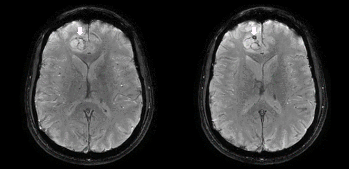
T2 gradient echo sequence versus susceptibility-weighted angiography  sequence in detecting microhemorrhages in hypertensive pati
T2 gradient echo sequence versus susceptibility-weighted angiography sequence in detecting microhemorrhages in hypertensive pati

Diagnosis of intracranial hemorrhagic lesions: comparison between 3D-SWAN  (3D T2*-weighted imaging with multi-echo acquisition) and 2D-T2*-weighted  imaging - Yoshiko Hayashida, Shingo Kakeda, Yasuhiro Hiai, Satoshi Ide,  Atsushi Ogasawara, Hodaka Ooki ...
Diagnosis of intracranial hemorrhagic lesions: comparison between 3D-SWAN (3D T2*-weighted imaging with multi-echo acquisition) and 2D-T2*-weighted imaging - Yoshiko Hayashida, Shingo Kakeda, Yasuhiro Hiai, Satoshi Ide, Atsushi Ogasawara, Hodaka Ooki ...

SWAN-Venule: An Optimized MRI Technique to Detect the Central Vein Sign in  MS Plaques | American Journal of Neuroradiology
SWAN-Venule: An Optimized MRI Technique to Detect the Central Vein Sign in MS Plaques | American Journal of Neuroradiology

Susceptibility-weighted angiography (SWAN) of cerebral veins and arteries  compared to TOF-MRA
Susceptibility-weighted angiography (SWAN) of cerebral veins and arteries compared to TOF-MRA

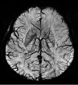
SWAN MR Application | GE HealthCare (United States)

MRI Technologist - MR Imaging of the brain in a patient with glioblastoma  multiforme (GBM). High resolution anatomical sequences (volumetric T2,  volumetric FLAIR, volumetric T1), 3D SWAN (for hemorrhage and vasculature  assessment)
MRI Technologist - MR Imaging of the brain in a patient with glioblastoma multiforme (GBM). High resolution anatomical sequences (volumetric T2, volumetric FLAIR, volumetric T1), 3D SWAN (for hemorrhage and vasculature assessment)
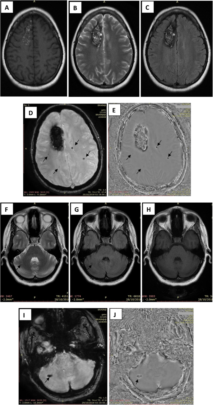
Magnetic resonance susceptibility weighted in evaluation of cerebrovascular  diseases | Egyptian Journal of Radiology and Nuclear Medicine | Full Text
Magnetic resonance susceptibility weighted in evaluation of cerebrovascular diseases | Egyptian Journal of Radiology and Nuclear Medicine | Full Text

GE Healthcare See brain vasculature in exquisite detail in one quick, easy  scan.
GE Healthcare See brain vasculature in exquisite detail in one quick, easy scan.

Figure 2 from Diagnosis of intracranial hemorrhagic lesions: comparison  between 3D-SWAN (3D T2*-weighted imaging with multi-echo acquisition) and  2D-T2*-weighted imaging | Semantic Scholar
Figure 2 from Diagnosis of intracranial hemorrhagic lesions: comparison between 3D-SWAN (3D T2*-weighted imaging with multi-echo acquisition) and 2D-T2*-weighted imaging | Semantic Scholar
Magnetic Resonance Imaging (MRI) | Radiology Key

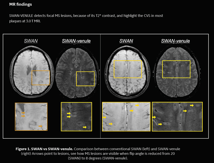
A new optimized 3T SWAN technique detects the central vein sign in MS  plaques in the clinical setting - Diagnóstico Journal
A new optimized 3T SWAN technique detects the central vein sign in MS plaques in the clinical setting - Diagnóstico Journal

EPOS™
EPOS™

Susceptibility-weighted Imaging: Technical Essentials and Clinical  Neurologic Applications | Radiology
Susceptibility-weighted Imaging: Technical Essentials and Clinical Neurologic Applications | Radiology
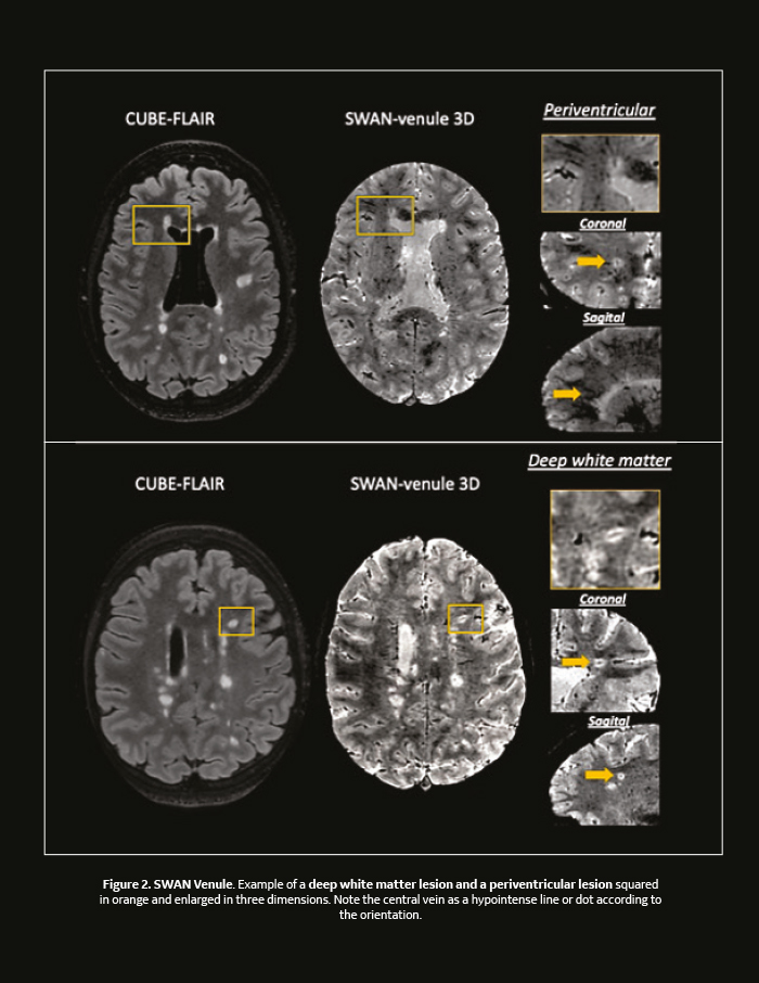
A new optimized 3T SWAN technique detects the central vein sign in MS  plaques in the clinical setting - Diagnóstico Journal
A new optimized 3T SWAN technique detects the central vein sign in MS plaques in the clinical setting - Diagnóstico Journal

SWI, susceptibiltiy - Questions and Answers ​in MRI

SWAN Brain Imaging by KMH Labs - Issuu

MRI (SWAN sequence: 3D SWAN TR: 79.3 msec, TE: 50.0 msec) Linear areas... |  Download Scientific Diagram
MRI (SWAN sequence: 3D SWAN TR: 79.3 msec, TE: 50.0 msec) Linear areas... | Download Scientific Diagram

SWI - Siemens Healthineers

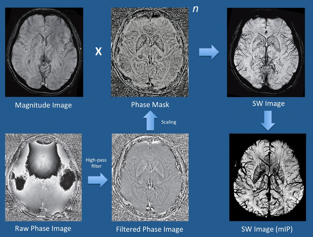
Susceptibility weighted imaging - Wikipedia

SWAN-Venule: An Optimized MRI Technique to Detect the Central Vein Sign in  MS Plaques | American Journal of Neuroradiology
SWAN-Venule: An Optimized MRI Technique to Detect the Central Vein Sign in MS Plaques | American Journal of Neuroradiology

Superficial haemosiderosis. GRE sequence (A) and SWAN (B). A sequel of... |  Download Scientific Diagram
Superficial haemosiderosis. GRE sequence (A) and SWAN (B). A sequel of... | Download Scientific Diagram

PDF] SWAN sequence in comparison to T2 for STN visualisation in DBS surgery  | Semantic Scholar
PDF] SWAN sequence in comparison to T2 for STN visualisation in DBS surgery | Semantic Scholar